National Institutes of Health Director Francis Collins, MD, PhD, wrote his blog this week about stunning SARS-CoV-2 images taken by the lab of Camille Ehre, PhD, at the UNC School of Medicine and the UNC Marsico Lung Institute.
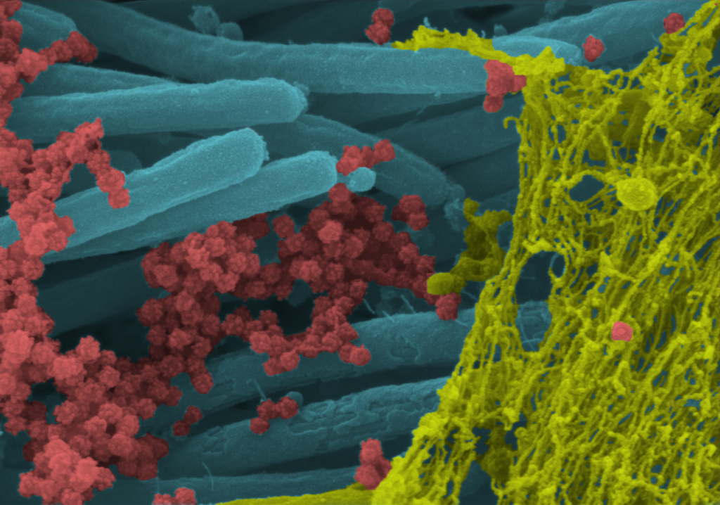
“If you or a loved one have come down with SARS-CoV-2, the coronavirus responsible for COVID-19, you know it often takes hold in the respiratory system. This image offers a striking example of exactly what happens to cells in the human airway when this coronavirus infects them.
This colorized scanning electron microscope (SEM) image shows SARS-CoV-2-infected human lung cells (purple) covered in hair-like cilia (blue). Those cilia line the inner surface of the airways and help to clear mucus (yellow-green) containing dust and other debris from the lungs. Emerging from the surface of those infected airway cells are many thousands of coronavirus particles (red).”
Read the full post from Dr. Collins here.
For more information on this work form Dr. Ehre and colleagues at the UNC Marsico Lung Institute, you can find it here.