In honor of University Research Week at UNC-Chapel Hill, the UNC School of Medicine is featuring three winning images from its “Art in Science” competition, images that embody the spirit of discovery, the importance of innovation and collaboration, and the visual beauty of biomedical research.

In celebration of University Research Week this week, the UNC School of Medicine Office of Research held an “Art in Science” competition, soliciting nominations for innovative scientific images that showcase the outstanding biomedical research in the school and the beauty that is reflected in the science.
More than 20 submissions were received, and after the images were evaluated for their aesthetic excellence, scientific or technical interest, and for their universal appeal for both scientists and non-scientists, three winning images were selected.
“Expecting a Special Delivery” was submitted by Kathleen Caron, PhD, Chair of the Department of Cell Biology and Physiology and member of the UNC Lineberger Comprehensive Cancer Center, Brooke Matson, PhD, and Kelsey Quinn, PhD, also from the cell biology and physiology.
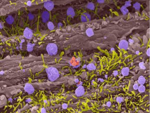 Like a lighted runway guiding the arrival of a plane with precious cargo, the uterus undergoes a dramatic transformation to create an ideal environment for an embryo to implant. For a brief window of time, cells on the surface of the uterus display spherical protrusions called pinopodes, which are thought to help guide and dock the incoming embryo. Several clinical studies in humans, as well as our own pre-clinical work in animal models, have found that pinopodes correlate with pregnancy success. However, the basic biology of pinopodes is elusive, and questions remain about how pinopodes support pregnancy. Pictured here is a scanning electron micrograph of several cells in a mouse uterus with pinopodes (violet) surrounded by microvilli (yellow); the central pinopode is secreting spherical structures (pink) that may aid in implantation. Further studies on pinopodes will reveal ways to improve fertility in women.
“Developing White Matter Pathways in the Infant Brain” was submitted by John Gilmore, MD, the Thad and Alice Eure Distinguished Professor and Vice Chair for Research and Scientific Affairs in the Department of Psychiatry and Jessica Girault, PhD, from the Department of Psychiatry.
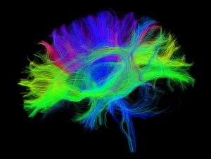 White matter fiber bundles in the human brain are organized during gestation and mature rapidly following birth. Magnetic resonance imaging, or MRI, is the primary method used for studying the developing human brain in-vivo. Here we employ a special type of MRI, called diffusion MRI, which captures diffusion patterns of water molecules in brain tissue, to trace bundles of axons connecting different regions in the brain. Using this method, we can reconstruct major fiber pathways in the brain. Quantitative information regarding the integrity of the axon bundles can be generated from these images and related to outcomes of interest in pediatric samples including cognition, language and motor abilities. This image shows the major fiber pathways reconstructed from an MRI of a 1-year-old infant; colors represent the primary orientation of the fiber bundles: green = anterior/posterior, red = left/right, blue = dorsal/ventral.
“Triple Connections” was submitted by Selena Romero, Matt Geden, PhD, and Mohanish Deshmukh, PhD professor of cell biology and physiology and member of the UNC Neuroscience Center and the UNC Lineberger Comprehensive Cancer Center.
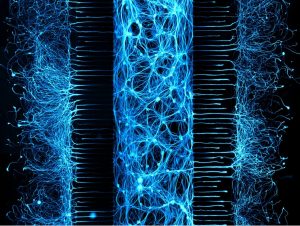 Neurons create stunning and complex networks needed to carry out everyday functions. This is accomplished by the neurons extending long processes, called axons, which reach out in search of distal targets. While the neuronal connections within the brain are remarkable, neurons isolated from the brain and maintained in a dish are just as capable of creating intricate and beautiful connections. This is demonstrated by this fluorescent micrograph depicting neurons that have been grown in a three chambered microfluidic device. Neuronal cell bodies plated in the center compartment extend their axons through microgrooves into outer compartments, creating three distinct and separated compartments. By using microfluidic devices such as this, we can assess how neurons regulate axonal health and how this affects overall neuron survival.
These stunning images are on display in Bondurant Hall lobby from November 4 – November 8. Congratulations to our winners!