During University Research Week, the labs of Dirk Dittmer, PhD, Jack Griffith, PhD, and Joshua Strauss, PhD, won the Art-in-Science Competition held by the UNC School of Medicine Office of Research.
Sponsored by the Office of the Vice Chancellor for Research at UNC-Chapel Hill, University Research Week featured speakers, presentations, and events, including the Art in Science Competition held by the School of Medicine Office of Research. Here are the winners:
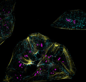
Allaura Cone, PhD, Meredith Chambers, and Dirk Dittmer, PhD, in the UNC Department of Microbiology & Immunology and the UNC Lineberger Comprehensive Cancer Center, were recognized for their image titled, “Endolysosomal Compartments in U-2 OS Cells,” fixed and stained with CD81 (cyan), LAMP1 (magenta), and Rhodamine-Phalloidin (yellow). Imaged at 100x on a confocal microscope, tetraspanin CD81 and Lysosome-associated membraine glycoprotein 1 (LAMP1) are important for the trafficking of extracellular vesicles throughout the cell.
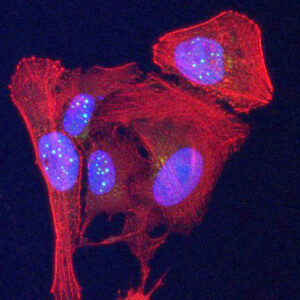 Taghreed Al-Turki, PhD, and Jack Griffith, PhD, in the Department of Microbiology & Immunology and the UNC Linberger Comprehensive Cancer Center, were recognized for their image titled “BRACO19 Disruption of Telomeres in U-2 OS Cells.” The image shows the cells in which the actine networks are stained red and the DNA in the nuclei stained blue. They used an antibody to detect the presence of a newly discovered protein, repeating arginine-valine dipeptide repeat protein expressed from the telomeres, and this is revealed as the light blue balls and particles in the nuclei. The buildup of this telomere protein was the result of treating the cells with an anti-cancer drug, BRACO19, which had been shown to specifically disrupt telomeres in cancer cells. Griffith holds a joint appointment in the UNC Department of Biochemistry & Biophysics.
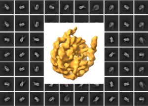 Emily Robinson and Joshua Strauss, PhD, in the UNC Department of Biochemistry & Biophysics and the Cryo-EM Core Facility, were recognized for their image titled, Nucleosome, a 3D reconstruction of a nucleosome against a backdrop of the initial 2D classes that came together to form the reconstruction. The nucleosome was imaged as part of the Cryo-EM Core’s development of novel lipid affinity grids.