A landmark study, led by Yen-Yu Ian Shih, PhD, professor of neurology and associate director of UNC’s Biomedical Research Imaging Center, could alter how researchers interpret results from functional magnetic resonance imaging.

The brain is an incredibly complex and active organ that uses electricity and chemicals to transmit and receive signals between its sub-regions. Researchers have explored various technologies to directly or indirectly measure these signals to learn more about the brain. Functional magnetic resonance imaging (fMRI), for example, allows them to detect brain activity via changes related to blood flow.
Yen-Yu Ian Shih, PhD, professor of neurology and associate director of UNC’s Biomedical Research Imaging Center, and his fellow lab members have long been curious about how neurochemicals in the brain regulate and influence neural activity, blood flow, and subsequently, fMRI measurement in the brain.
A new study by the lab has confirmed their suspicions that fMRI interpretation is not as straightforward as it seems.
“Neurochemical signaling to blood vessels is less frequently considered when interpreting fMRI data,” said Shih, who also leads the Center for Animal MRI. “In our study on rodent models, we showed that neurochemicals, aside from their well-known signaling actions to typical brain cells, also signal to blood vessels, and this could have significant contributions to fMRI measurements.”
Their findings, published in Nature Communications, stem from a $3.8-million grant from the National Institutes of Health and UNC’s investments in supporting the installation and upgrade of two 9.4-Tesla animal MRI systems and a 7-Tesla human MRI system at the Biomedical Research Imaging Center.
When activity in neurons increases in a specific brain region, blood flow and oxygen levels increase in the area, usually proportionate to the strength of neural activity. Researchers decided to use this phenomenon to their advantage and eventually developed fMRI techniques to detect these changes in the brain.
For years, this method has helped researchers better understand brain function and influenced their knowledge about human cognition and behavior. The new study from Shih’s lab, however, demonstrates that this well-established neuro-vascular relationship does not apply across the entire brain because cell types and neurochemicals vary across brain areas.
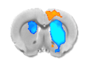
Shih’s team focused on the striatum, a region deep in the brain involved in cognition, motivation, reward, and sensorimotor function, to identify the ways in which certain neurochemicals and cell types in the brain region may be influencing fMRI signals.
For their study, Shih’s lab controlled neural activity in rodent brains using a light-based technique, while measuring electrical, optical, chemical, and vascular signals to help interpret fMRI data. The researchers then manipulated the brain’s chemical signaling by injecting different drugs into the brain and evaluated how the drugs influenced the fMRI responses.
They found that in some cases, neural activity in the striatum went up, but the blood vessels constricted, causing negative fMRI signals. This is related to internal opioid signaling in the striatum. Conversely, when another neurochemical, dopamine, predominated signaling in striatum, the fMRI signals were positive.
“We identified several instances where fMRI signals in the striatum can look quite different from expected,” said Shih. “It’s important to be mindful of underlying neurochemical signaling that can influence blood vessels or perivascular cells in parallel, potentially overshadowing the fMRI signal changes triggered by neural activity.”
Members of Shih’s lab, including first- and co-authors Dominic Cerri, PhD, and Lindsey Walton, PhD, travelled to the University of Sussex in the United Kingdom, where they were able to perform experiments and further demonstrate the opioid’s vascular effects.
They also collected human fMRI data at UNC’s 7-Tesla MRI system and collaborated with researchers at Stanford University to explore possible findings using transcranial magnetic stimulation, a procedure that uses magnetic fields to stimulate the human brain.
By better understanding fMRI signaling, basic science researchers and physician scientists will be able to provide more precise insights into neural activity changes in healthy brains, as well as in cases of neurological and neuropsychiatric disorders.
The research was supported by Brain Research through Advancing Innovative Neurotechnologies (BRAIN) Initiative grant RF1MH117053 and High-End Instrumentation grants S10OD026796 and S10MH124745from the National Institutes of Health.
Media contact: Kendall Daniels, Communications Specialist, UNC Health | UNC School of Medicine