In honor of University Research Week, the UNC School of Medicine is featuring three winning images from its “Art in Science” competition, images that embody the spirit of discovery, the importance of innovation and collaboration, and the visual beauty of biomedical research.
In celebration of University Research Week this week, the UNC School of Medicine Office of Research held an “Art in Science” competition, soliciting nominations for innovative scientific images that showcase the outstanding biomedical research in the school and the beauty that is reflected in the science. This is the second year that the competition has been held.
After all submissions were evaluated for their aesthetic excellence, scientific or technical interest, and for their universal appeal for both scientists and non-scientists, three winning images were selected.
The following winning images will be on display in Bondurant Hall from November 8 – 12.
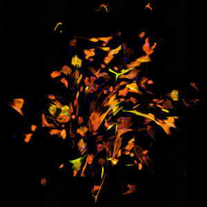
“Eclipsed” was submitted by Benjamin Keepers and Dr. Li Qian from the Department of Pathology and Laboratory Medicine. This image was stitched together from dozens of fluorescence microscopy images. It shows a culture of human fibroblasts which have been reprogrammed into cardiomyocyte-like cells. The researchers performed immunofluorescence to allow for visualization of different molecules in the cell: DNA is stained in blue; cardiac Troponin T and αActinin – two cardiomyocyte-specific markers – are stained in orange and green, respectively. A majority of the cells in this sample have been reprogrammed, and they express these cardiac markers. This was a surprising result for the team – because the boost in reprogramming efficiency came not from a cardiac factor, but a neuron factor, suggesting that this neuron factor can “moonlight” in cardiac reprogramming.
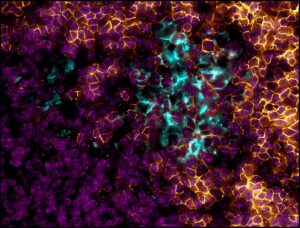
“Hotspot of Immune Response to SARS-CoV-2 Spike Protein” was submitted by Anthony Brent Eason and Dr. Dirk Dittmer from the Department of Microbiology and Immunology.
In a mouse spleen, immune cells react to SARS-CoV-2 spike protein immunization by making antibodies against the virus. The germinal center contains activated B cells, which may produce protective antibodies. B cells are shown in gold and activated B cells (the germinal center) are colored cyan. Each cell in the spleen has a nucleus containing DNA, shown in magenta. The team uses findings from mouse trials to develop more efficient, robust, and safe vaccine candidates and delivery systems for humans.
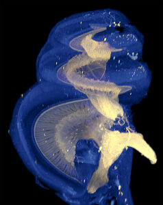
“The Cochlea” was submitted by Drs. Ken Hutson and Doug Fitzpatrick from the Department of Otolaryngology-Head and Neck Surgery. This is a three-dimensional image of a gerbil’s cochlea, the end organ for hearing, taken with the LaVision II fluorescent light sheet microscope in the Microscopy Services Laboratory Core Facility (Director, Dr. Pablo Ariel). Spiral ganglion cells – neurons that receive auditory information through their fan-like peripheral processes – transmit this information to the brain through the auditory nerve. These elements have been immunolabeled with neuron-specific beta-tubulin (yellow). Other structures visible through autofluorescence (blue) include hair cells and the stria vascularis.