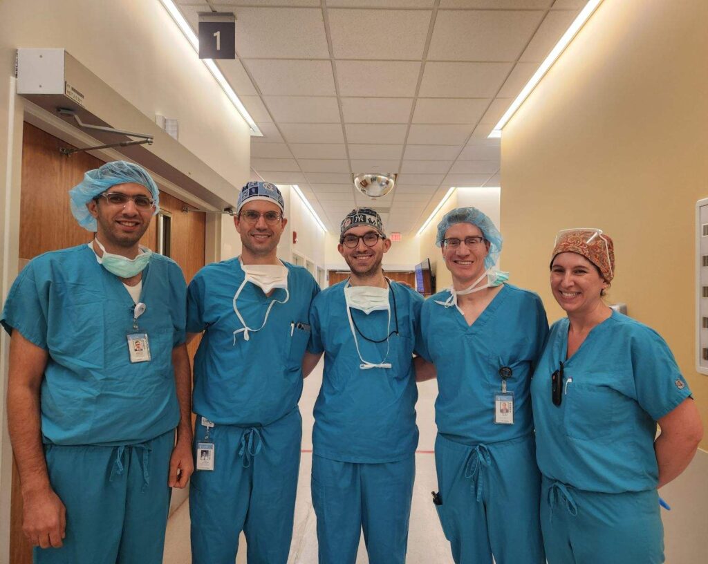
Matthew Miller, MD, Daniel Rubinstein, MD, and Hussam Banna, MD, from the UNC School of Medicine joined forces to perform the first corneal neurotization procedure at UNC, a life-changing surgery for patients who have neurotrophic keratitis.
For the first time at UNC Hospitals, a surgical team has successfully performed corneal neurotization — the only disease modifying surgical treatment for neurotrophic keratitis (NK), a rare eye condition that causes loss of sensation to the cornea and can lead to permanent vision loss, if left untreated.
“It’s a life-changing procedure,” said Matthew Miller, MD, director of the UNC Facial Nerve Center and assistant professor of otolaryngology/head and neck surgery at the UNC School of Medicine. “It not only restores sensation in their eyes, but it can prevent and even reverse vision loss.”
This procedure fills an important access gap, as the UNC team is the only one performing the surgery in the states of North Carolina, South Carolina, and Virginia. There are just 20 to 30 groups performing corneal neurotization in the United States.
What is Neurotrophic Keratitis?
The cornea plays a few pivotal roles for the overall health of the eyeball. Much like the windshield system of a car, the cornea helps protect the inner structures of the eye and keeps the surface of the eye clean and moist. When debris lands on the surface or it begins to dry out, the cornea triggers the blink reflex.
Blinking helps to moisturize the eye and sweeps off harmful debris and bacteria. The cornea is also a critical component of the optical system of the eye, and its clarity and regularity of its surface are vital to proper visual function.
Neurotrophic keratitis, which affects about 5,000 to 10,000 people in the United States, occurs when the nerves innervating the cornea are damaged. The most common causes of NK are trauma to the eye, infectious diseases, brain tumors, and brain surgery. With its nerve supply cut off, the cornea is unable to keep itself moist or maintain a healthy architecture, causing ulceration, scarring, and eventually a permanent loss of vision.
“If you lose your blink reflex, your cornea can become unhealthy, dry, and very inflamed,” said Daniel Rubinstein, MD, assistant professor of ophthalmology and oculofacial plastic surgeon on the case. “But you can’t feel the pain or dryness. Over time, the outermost layer of the cornea can slough off and cause ulceration and scarring, which impairs your vision.”
In the most severe of cases, the affected eye needs to be removed to prevent widespread infection and further damage to other structures.
How Does Corneal Neurotization Work?
Simply put, the goal of the surgery is to transfer a nearby healthy nerve to the damaged cornea to restore its sensation.
During the procedure, Miller harvests a segment of sural nerve from the lower leg. This will be used as a graft, or bridge, to connect a healthy nerve to the damaged cornea. Surgeons prefer to harvest this nerve because its sacrifice leaves patients with only a small area of numbness on top of the foot.
Rubinstein then make a small incision in each upper eyelid and accesses the heathy supraorbital nerve just beneath the eyebrow. He then tunnels the sural nerve graft through the incisions across the upper face to connect the affected eye to the healthy supraorbital nerve.
Using a specialized microscope, Miller carefully peels back the individual nerve fibers that make up the sural nerve. Hussam Banna, MD, assistant professor of ophthalmology and cornea specialist, then sews the fibers to the damaged cornea itself. Miller completes the circuit by sewing the other end of the sural nerve to a healthy “donor” nerve under the microscope.
After approximately six months, the patient may start to notice improved sensation and, ultimately, vision. The healing process is slow, but life changing for patients.
According to Miller, the minimally invasive nature of the surgery is especially important for patients because it reduces scarring on the face, numbness, and other potential side effects.
“It is important because these patients, especially patients with facial paralysis and neurotrophic keratitis, have gone through very traumatic events,” said Miller. “Our faces are our daily life. The last thing you want to do is create an overly visible scar to create another reminder of the traumatic experiences of their past.”
A Procedure Unique to UNC

Performing the complex procedure requires tight-knit collaboration among operating room nurses, technical staff, coordinators, and, in this case, three surgeons.
Experts in their own rights, Miller, Rubinstein, and Banna, united forces at UNC Hospitals campus at Hillsborough, which housed all of the specific medical instruments needed for the surgery.
“It’s pretty rare to have three surgeons operating on the same case, coordinating, and simultaneously taking care of the same patient,” said Rubinstein. “This kind of cooperative care for a patient is rather unique to UNC. We are incredibly grateful to have received so much support from the hospital system and the institution to perform such an innovative, life-changing procedure for these patients. We look forward to continuing our collaboration to offer this treatment to any patient that might benefit.”
Media contact: Kendall Daniels, Communications Specialist, UNC Health | UNC School of Medicine